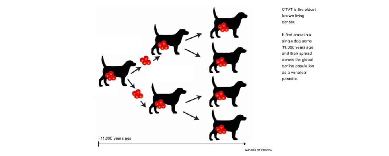
By Dr Abdul Mateen and Syed Balaj Hussain Rizvi, College of Veterinary and Animal Sciences, University of Veterinary and Animal Sciences, Jhang “It is a histiocytic tumor of the external genitalia of the dog and other canines, and is transmitted from animal to animal during mating”.
Other Names include Transmissible Venereal Tumours (TVTs), canine transmissible venereal sarcoma (CTVS), infectious or venereal granuloma, Sticker tumour, transmissible sarcoma, and contagious venereal tumour and infectious sarcoma.
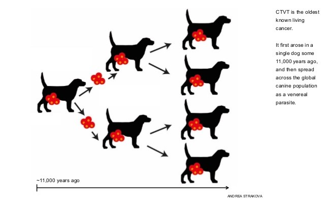
History: CTVT first emerged in a dog that lived about 11,000 years ago. All CTVT tumours carry the DNA belonging to this “founder dog”. By counting and analysing the mutations acquired by CTVT tumours worldwide, we can piece together how and when CTVT emerged and spread. CTVT is thus the oldest cancer known in nature.
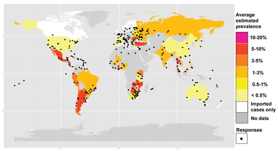
Epidemiology:CTVT is a common disease in dogs around the world. Its global distribution is associated with the presence of free-roaming dogs. In the United Kingdom, CTVT disappeared during the twentieth century as an unintentional result of the introduction of dog control laws.
Aetiology:A virus or virus-like organism does not cause it. The canine transmissible venereal tumour’s infectious agent is the cancer cell itself, and the tumour is clonal in origin.
CTVT CYCLE:
CTVT undergoes a predictable cycle:
- The initial growth phase of 4–6 months (P phase)Higher mast cell counts, High microvessel counts & P‐phase tumours consist of round cells with microvilli
- Stable phase (S phase)It consists of cells undergoing the transition from round cells to spindle‐shaped fibroblasts
- Regression phase (R phase) Although not all tumours will regress. Regressing tumours contain higher numbers of lymphocytes, most of which are T cells
Transmission:Transmission can occur through one of the following methods.
- Horizontal transmission from one dog to another dog
- Direct contact with tumour cells
- Sexual intercourse with diseased animal
- By oral contact with diseased part (i.e. sniffing and licking of the affected area)
- Transmission from the dam to the pup during social interactions such as grooming and other maternal behaviour
Sites of Infection:The most common primary sites of involvement are the external genitalia. Other locations can be affected on occasion, including the nasal and oral cavities, subcutaneous tissues, and the eyes.
Sign and SymptomsSign & symptoms of CTVT includes
In male dogs, the tumour affects
- the penis and foreskin
- In female dogs, it affects the vulva
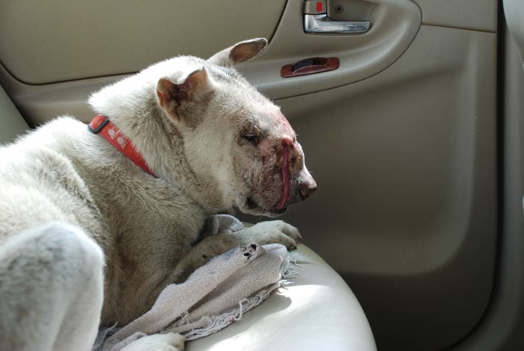
- Rarely, the mouth or nose are affected
- The tumour often has a cauliflower-like appearance
- A red, tumorous mass was bulging out of the surface membrane of the vagina or on the penis.
- Blood drops may also be observed dripping from the vagina or penile foreskin.
The dog will usually lick the affected area with frequency.
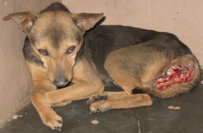
Signs of genital TVT include
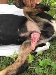
- A discharge from the prepuce
- In some cases urinary retention, from blockage of the urethra.
Signs of a nasal TVT include
- Nasal fistulae
- Nose bleeds
- Other nasal discharge
- Facial swelling
- Enlargement of the sub-mandibular lymph nodes
 Diagnosis:A complete health history, with as much information as possible regarding when the symptoms began, how much freedom your dog has to roam freely or whether other dogs roam the area if you have been attempting to breed your dog, etc. The physical examination will focus primarily on your dog’s genital organs. A tissue sample of the mass will need to be taken for biopsy, and fluid samples will be taken for the standard laboratory tests, including the complete blood count, biochemistry profile and urinalysis. These tests are usually within normal ranges, but in some cases, blood cells or abnormal cancer cells may be found in the urine sample. This type of tumour rarely spreads to other locations, but your doctor will need to visually confirm that it is not a malignant form of cancer. Visual diagnostics will include chest and abdominal X-rays to determine if there has been metastasis and its stage if cancer is present. Your veterinarian will also palpate the affected area’s lymph nodes to determine how much the lymph nodes are reacting to the abnormality, an important distinguishing factor when cancerous cells are present. A sample of lymph fluid will be sent to a laboratory for further evaluation to determine if cancerous cells are in the sample. The presence of cancerous cells in the lymph nodes is often a strong indication that the tumour is not benign. Treatment will be based on the results of these tests.
Treatment: Transmissible venereal tumours are usually progressive and are treated with a combination of:
Surgical technique: removal of the tumour through an en-bloc technique
Chemotherapy: intravenous administration of vincristine sulphate at the dose of 0.025 mg kg−1 of body weight, once a week, for four weeks or Doxorubicin HCL can also be used 30mg per m2of the lesion with the maximum cumulative dose of 240mg/m2.


